Email Address
info@ultrasound-scanners.co.uk
Phone Number
+44(0) 7860 637522
Our Location
London — Nottingham
info@ultrasound-scanners.co.uk
+44(0) 7860 637522
London — Nottingham
yiannis
December 21, 2024

Ultrasound is sound waves with frequencies higher than the upper audible limit of human hearing. Ultrasound is not different from the “normal” (audible) sound in its physical properties, except that humans cannot hear it. This limit varies from person to person, and it is approximately 20 kilohertz (20,000 hertz) in young adults.
Sonar (originally an acronym for sound navigation ranging) is a technique that uses ultrasound to detect, locate, and determine the size and shape of underwater objects, complemented by techniques using electromagnetic waves such as radar and ladar.
Medical ultrasound is used to visualize muscles, tendons, and ligaments; examine the abdomen, pelvis, and prenatal foetus, and guide procedures such as needle biopsies and aspirations.
Ultrasound can also be used to measure blood flow and investigate heart function. Because ultrasound waves can be transmitted through fluid-filled tissues with no reflections, this technology is ideal for examining babies in the womb.
In addition to its medical uses, ultrasound has found a place in a variety of other industries. For example, it is used in beauty salons for cosmetic treatments like facial toning and cellulite reduction. Ultrasonic cleaning equipment is also used in many industrial settings to clean delicate machinery parts or remove contaminants from surfaces.
Ultrasound waves were first discovered in 1794 by Christian Johann Daniel von Recklinghausen, a German physicist. However, it was not until the early 1900s that medical professionals began to realize the potential of using ultrasound waves for diagnostic purposes. In the 1930s, researchers in both the United States and Europe began to experiment with using ultrasound for therapeutic purposes.
The use of ultrasound in medicine started in the early 1800s with Jean-Daniel Colladon, a Swiss physicist, successfully using an underwater bell to determine the speed of sound in the waters of Lake Geneva and in 1877 Lord Rayleigh in England publised “the Theory of Sound” which first described sound wave as a mathematical equation, forming the basis of future practical work in acoustics.
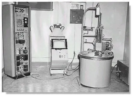
Dr. Karl Theodore Dussik in Austria in 1942, published the first on medical ultrasonics. The work of Professor Ian Donald and his colleagues in Glasgow in the mid-1950s helped to facilitate the development of practical techniques and applications in medical ultrasound. Since then, ultrasound technology has progressed rapidly and accelerated by the use of computers. These days, ultrasound is being used to image most of the body organs, and it is the investigation of choice in pregnancy.
The history of medical ultrasound began with SONAR (Sound Navigation and Ranging) for submarines and has had many uses with varying degrees of success since then.
Ultrasound history, medically speaking, has been primarily a diagnostic technology, although it has been tested and used for therapy as well. Doctors and sonographers have been capturing images from within the human body since the 1940s and in spite of its varied history, ultrasound has become one of the most widely used medical diagnostic tools in modern medicine.
The use of ultrasound in medicine started in the early 1800s with Jean-Daniel Colladon, a Swiss physicist, successfully using an underwater bell to determine the speed of sound in the waters of Lake Geneva and in 1877 Lord Rayleigh in England publised “the Theory of Sound” which first described sound wave as a mathematical equation, forming the basis of future practical work in acoustics.
Dr. Karl Theodore Dussik in Austria in 1942, published the first on medical ultrasonics. The work of Professor Ian Donald and his colleagues in Glasgow in the mid-1950s helped to facilitate the development of practical techniques and applications in medical ultrasound. Since then, ultrasound technology has progressed rapidly and accelerated by the use of computers. These days, ultrasound is being used to image most of the body organs, and it is the investigation of choice in pregnancy.
.
Ultrasound is a high-frequency sound, typically above 20 kilohertz (kHz). The upper-frequency limit of human hearing is around 20 kHz. Sound above this level is called ultrasound. Humans with normal hearing can sometimes hear ultrasound, especially if it is pulses of sound with a low-duty cycle (i.e., high intensity and low repetition rate).
The pitch of the sound that humans can hear depends on its frequency. The pitch of ultrasound is thus too high for humans to hear. However, some animals, such as bats, can produce and receive an ultrasound for echolocation.
The reflectivity of ultrasound also depends on its frequency. For medical imaging, frequencies between 2 and 20 MHz are typically used. At these frequencies, most soft tissues are relatively Reflective, while bone and air are relatively absorbent.
Ultrasound waves are generated by piezoelectric transducers. These transducers convert electrical energy into ultrasound waves or vice versa. The waves travel through the body and bounce off tissues in a process known as scattering. When the waves hit something that they cannot pass through, such as a bone, they reflect towards the transducer. By measuring the time it takes for the reflected waves to return, it is possible to construct an image of the internal organs using a process known as echolocation .
Ultrasound has become an important tool in modern medicine. It is used in a variety of medical fields, such as obstetrics, cardiology, and neurology. With the advancement of technology, ultrasound is only going to become more important in the medical field.
In the past few years, there has been a dramatic increase in research of the use of ultrasound for a variety of new applications. These include:
-Using ultrasound to treat cancerous tumours
-Using ultrasound to dissolve kidney stones
-Using ultrasound to treat Alzheimer’s disease
-Using ultrasound to improve the efficacy of chemotherapy drugs
– using ultrasound to deliver drugs directly to cancerous tumours
– using ultrasound to image the brain in real time
Independent supplier of bladder scanners and new and used ultrasound scanners for sale.

Ultrasound-Scanners.co.uk
Copyright © 2025. All rights reserved.