Cart is empty.

info@ultrasound-scanners.co.uk
+44(0) 7860 637522
London — Nottingham
yiannis
December 21, 2024
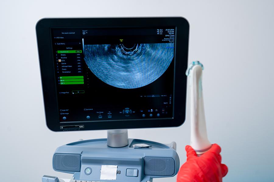
Current diagnostic ultrasonography technology uses probes containing an acoustic ultrasound transducer (or several ultrasound transducers) to send pulses of ultrasound into matter. When the mechanical wave comes upon a boundary between two different materials (different acoustic impedance) energy is reflected back to the ultrasound transducer. This reflected energy echo is detected by theprobe. The time it takes for the echo to travel back to the probe is electronically measured and used to calculate, as well as to display, the depth of the tissue.
The frequencies used for medical ultrasonography are generally in the range of 1 to 15 MHz. Higher frequencies have a lower wavelength, producing images with a greater resolution. However, the reduction of the wave is increased at higher frequencies, so in order to most effectively penetrate deeper tissues, a lower frequency (3-5MHz) is used.
Ultrasound transducers produce images of muscle and soft tissue, useful for defining borders between solid and fluid-filled spaces. Ultrasound transducers provide live images, enabling operators to select the most useful sections for rapid diagnoses. They show the structure and function of internal organs. It provide a useful way to examine the musculoskeletal system to detect problems with muscles, ligaments, tendons and joints. It also assist in identifying blockages, stenosis and other vascular abnormalities.
Modern, high-class ultrasoundsystems use the best ultrasound transducers technology, coupled with excellent processors and a user friendly interface. The image quality depends mainly on the ultrasound transducer, which is the front end that transmits and receives the mechanical energy. Modern ultrasound transducers give users unparalled and unsurpassed multi-modality ultrasound experience.
The most common ultrasound applications are:
•Portable ultrasound machines
•OB/GYN ultrasound
•Cardiac ultrasound
•Abdominal ultrasound
•Urology ultrasound
•Vascular ultrasound
•3D/4D ultrasound scanners
•Musculoskeletal ultrasound
•Transcranial Doppler systems
•Small particles scanners
•Ophthalmology
•Veterinary ultrasound machines
The most popular types of ultrasound are:
•Linear probes
•Phased array probes
•Convex ultrasound probes
•Real time 4D ultrasound probes
•Endocavity ultrasound probes
•Endovaginal ultrasound probes
•Laparoscopic ultrasound probes
•Intravascular ultrasound probes
Other uses of ultrasound equipment.
•Carotid ultrasonography, used to assess blood flow into the carotid arteries, as well as the intra-cerebral arteries.
•Echocardiography, which is an ultrasound that shows the movement of the heart, as the muscle dilates and contracts.
•Emergency medical technicians often use a form of probe. Also, it is used in the ER as a routine method of quickly assessing the cause of a patient’s abdominal pain.
•Urologists often use ultrasound transducers to detect the level of fluid that is in a patient’s bladder.
•Gynecologists may perform a pelvic sonogram using an ultrasound to view an image of the pelvic floor in women and diagnose any abnormalities.
•More detailed images of the tendons, nerves, muscles, ligaments and other soft tissue areas that may have been affected by injury or trauma.
•Arterial probe is used by cardiologists to check for possible obstructions in the arteries, or to diagnose DVT.
•In gastroenterology, doctors can view the abdomen using an probe to view organs such as the aorta, pancreas, gall bladder, kidneys, liver and spleen.